M Mode Ultrasound Diaphragm

Diaphragm M Mode Ultrasound Imaging From An Anterior Subcostal

Classification Of Diaphragm Function By M Mode Ultrasound With The

M Mode Sonography For The Measurement Of Diaphragm Movement
Diaphragmatic Paralysis The Use Of M Mode Ultrasound For
Diaphragmatic Paralysis The Use Of M Mode Ultrasound For

A Probe Position For B And M Mode Diaphragmatic Excursion
And philippe blanc md background.
M mode ultrasound diaphragm. It can be performed if necessary at the. Ultrasound imaging is a noninvasive radiation free accurate and safe technique allowing assessment of diaphragm anatomy and function. The aim ofthis prospective study was to determine the reference values for diaphragmatic motion as recorded. Neuromuscular ultrasound of the diaphragm is an evolving diagnostic modality with several techniques and measurements that can be employed for structural and functional assessment of the diaphragm.
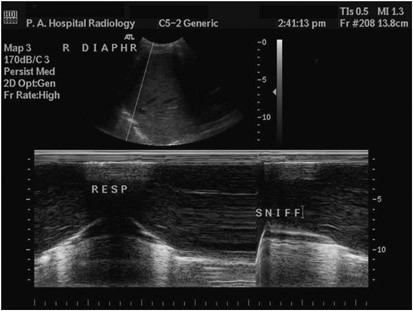
The authors review the pathophysiology of diaphragm in neuromuscular disorders the methodology and indications of diaphragm ultrasound imaging as well as possible pitfalls in the interpretation of results. M mode ultrasonography is a relatively simple and accurate test for diagnosing paralysis of the diaphragm in the adult population. For vs measurement the amplitude of excursion was measured on the vertical axis of the tracing from the baseline to. The m mode trace of the paralyzed side showed no active caudal movement of the diaphragm with inspiration and abnormal paradoxical movement ie cranial movement on inspiration particularly with the sniff test.
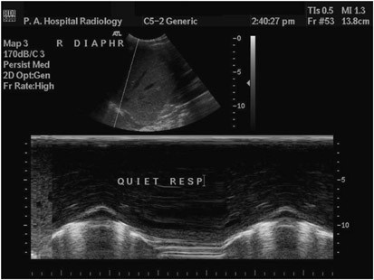
Imaging of the diaphragm dome using m mode with both quiet breathing and with performance of maneuvers such as a sniff test see below and the appropriate interpretation of diaphragm motion with these maneuvers should be viewed as more advanced competencies in diaphragm ultrasound. Using m mode the diaphragm is seen as a single thick echogenic line. Although diaphragmatic motion is readily studied by ultrasonography the proce dure remains poorly codified. M mode diaphragm assessment is ideal in an icu patient with acute respiratory failure as it enables the physician to understand whether or not diaphragm dysfunction is implicated in the acute respiratory failure early goal directed us examination.
In cases of unilateral diaphragmatic paralysis the affected side demonstrates a paradoxical upward movement. M mode ultrasound for evaluation of diaphragmatic movement abnormalities has previously been described most notably in the pediatric population 1 2 3 including a recent large series of 278. Identification of diaphragm. For the qb and db measurements the first caliper was placed at the foot of the inspiration slope on the diaphragm echoic line and the second caliper was placed at the apex of this slope.
The diaphragm inspiratory amplitudes excursions were measured from the m mode sonography. 19 it also can be used at the time of weaning from mv during a spontaneous breathing trial late application. Fluoroscopic examination of the diaphragm sniff test is very useful in diagnosing a diaphragmatic paralysis. 20 in contrast it is not useful if.
This is due to the technical challenges of image acquisition especially of the left hemidiaphragm and also.

Diaphragmatic Paralysis The Use Of M Mode Ultrasound For

Ultrasonographic M Mode Images From The Right Hemidiaphragm A
M Mode Sonography Of Diaphragmatic Excursion The Amplitude Of

Diaphragm In B And M Mode In Spontaneous Breathing A And In

Evaluation Of Diaphragmatic Thickness And Mobility A Example Of

Sonographic Evaluation Of Diaphragmatic Excursion And Thickness In

Sonography Of The Diaphragm In The Zone Of Apposition In B Mode

M Mode Sonography Of The Diaphragm Of A A Representative Patient

