M Mode Ultrasound Lung
M Mode For Pneumothorax Emory School Of Medicine

M Mode Ultrasound Of The Right Lung Demonstrating Bleb Point A

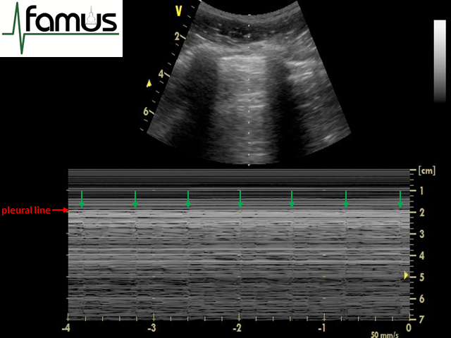
Barcode Sign In Pneumothorax On Pocus M Mode Seashore Sign

M Mode Ultrasound Images Demonstrating A The Appearance Of Lung
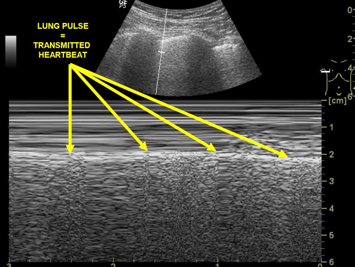
Lung Point On Motion Mode M Mode The Arrow Indicates A
Ultrasound For Detection Of Pneumothorax Rebel Em Emergency
One hundred forty emergency.
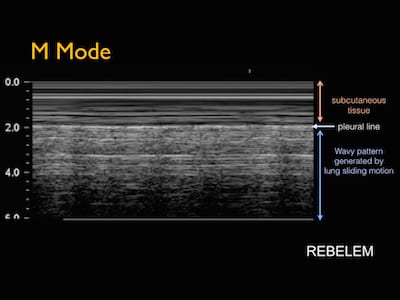
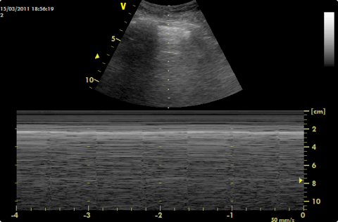
M mode ultrasound lung. Often utilized for its excellent axial and temporal resolution of structures m mode or motion mode is a form of ultrasonography in which a single scan line is emitted received and displayed graphically. The m mode takes a single line of echoes from the two dimensional image and plots it against time. Slide 3 of 3. Linear normal lung slide.
Pneumothorax m motion mode. Normal motion seen below pleural line seashore sign continue narration. Lung normal lung slide. Ants on a string.
Our study used m mode ultrasonography of the pleura to differentiate cpe from ncais. This study aimed to determine what effect if any this addition has on us interpretation by emergency physicians of varying training levels. Since lung sliding is sometimes challenging to visualize using the motion mode m mode on ultrasound can help to confirm findings. On the affected side we see a lines absence of pleural sli.
Plaps profile visceral pleura quoad sign 2d lus sinusoid sign. M mode ultrasound andrew murphy and dr david carroll et al. The a lines normal lung pleural line a lines a lines linear probe convex probe the a lines are horizontal artifactual repetitions of the pleural line displayed at regular intervals. Lung ultrasound versus chest radiography for the diagnosis of pneumothorax in critically ill patients.
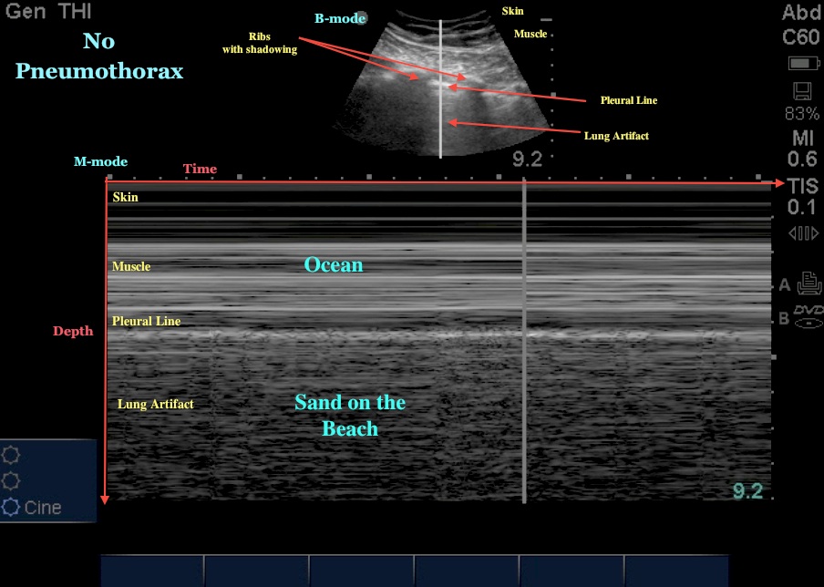
It is unknown whether the addition of m mode to b mode ultrasound us has any effect on the overall accuracy of interpretation of lung sliding in the evaluation of a pneumothorax by emergency physicians. On the unaffected side we see a lines pleural sliding in b mode and seashore sign in m mode. Often utilised for its excellent axial and temporal resolution of structures m mode or motion mode is a form of ultrasonography in which a single scan line is emitted received and displayed graphically. Slide 2 of 3.
Normal lung 2d m mode ribs pleural line anterior lung surface bat sign sea and sand sign sector probe. Slide 1 of 3. M mode ultrasound patrick o shea and dr david carroll et al. A prospective single blind study.
An m mode recording is conventionally displayed with the abscissa representing time and the ordinate distance from the. Below are side by side examples of normal pleural slide and pneumothorax depicted in both b mode and m mode with linear sector and curvilinear probes. An m mode recording is conventionally displayed with the abscissa representing time and the ordinate distance from the. The presence of b lines on lung ultrasonography is a characteristic feature of both cardiogenic pulmonary edema cpe and noncardiogenic alveolar interstitial syndrome ncais so their presence does not allow the clinician to differentiate between the two entities.
Normal lung slide. The manner in which the image is produced may be thought of as a graph with the x axis representing time and the y axis the depth when using m mode the screen.
2 Lung Ultrasound Pre Reading For Fcus Course Intensive Care Network
Pneumothorax

M Mode For Pneumothorax Lesson 87

Normal Lung Versus Pneumothorax M Mode Download Scientific

M Mode Doppler Ultrasound Demonstrating Discrimination Of A

M Mode Sonography For The Measurement Of Diaphragm Movement

Abnormal Lung Sliding M Mode Ultrasound Jetem 2018 Jetem

Normal Lung Sliding M Mode Ultrasound Jetem 2018 Jetem