Mitral Valve M Mode Points

M Mode Display Of Mitral Valve Anterior Mitral Valve Leaflet

Mitral Valve M Mode Echocardiogram Radiology Case

M Mode Labeling Mitral Valve Diagram Quizlet
Valves Thoracic Key
2 4 M Mode 123sonography

Elucidating The B Bump On The Mitral Valve M Mode Echogram In
M mode examination of the mitral valve the m mode examination of the mitral valve with respect to the cursor position structures interrogated and the normal motion of these structures is displayed in figure 4 2.
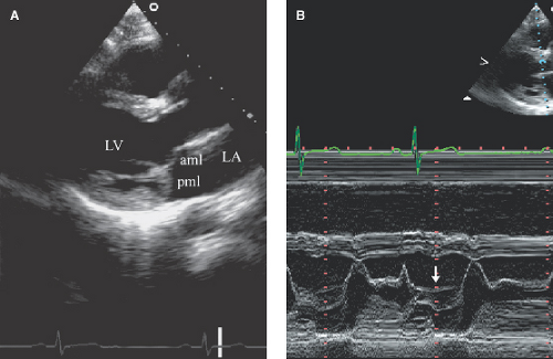
Mitral valve m mode points. 3d reconstructions identified the mitral valve annulus as a hyperbolic paraboloid or a saddle shaped structure with curved planes parallel to the anteroposterior axis opening upward and orthogonal curved planes that open downward. Holosystolic sagging back of the anterior posterior or both mv leaflet 3mm from the c d point of mv. One may appreciate the approximated mitral valve leaflets at the beginning of diastole d swing apart with the cephalad and anterior excursion of the anterior leaflet contacting the septum at its peak excursion e. As a double heart valve replacement patient i remember that one of the most critical steps in the diagnosis of my defective aortic heart valve was the echocardiogram.
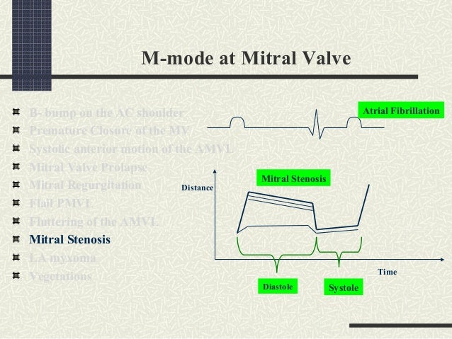
Mid to late systolic sagging back of the anterior posterior or both mv leaflet 2mm from c d point of mv. Brief explanation of m mode motion mode ultrasound. Ekg tracingt waveqrs complexp wavesystole diastole 12. M mode echocardiog raphy provides an ice pick one dimensional depth only view of the heart 1 2 the ultrasound echoes reflected from the various car diac interfaces are represented as dots and their intensities by.
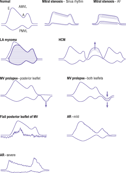
Mitral valve prolapse. M mode at the mitral valvethe mitral valve has 2 leaflets anterior andposterior specific letters corresponding to systole anddiastole are assigned to the m mode tracingof the mitral valve. Jude mitral prosthetic valve. Table 4 3 at the end of this chapter lists the abnormalities that may be detected from this m mode examination.
9 m mode echocardiography itzhak kronzon md gerard p. Gulfcoast ultrasound institute 88 491 views. Aurigemma md historically m mode motionmode wasthefirsteffectivemodal ity for the ultrasonic evaluation of the heart. As the pressure gradient between the left atrium and.
Distancesystolem mode at mitral valvediastoletime 13. Imaging plane and position of m mode cursor the mitral valve can be. M mode in recording motion of a disk type prosthetic valve m mode echocardiogram of a st. M mode features thick redundant mitral valve leaflets.
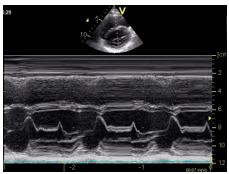
2 4 2 m mode at the level of the mitral valve this application of the m mode was very important in the early days of echocardiography prior to the discovery of two dimensional imaging. M mode echocardiography is ideal to record the brisk opening and closing of the disks arrows. An m mode echocardiogram from the parasternal long axis with the m mode pick directed through the mitral valve leaflets. 3de has increased the understanding of the anatomy of the mitral valve annulus more than that of any other structure in the heart.
It displays mitral valve motion and was mainly used to quantify mitral stenosis as well as visualize mitral valve prolapse and systolic anterior motion sam of the mitral valve in hypertrophic cardiomyopathy.
Mitral Regurgitation Anesthesia Key

M Mode At Center For Ultrasound Research And Education Studyblue
Http Www Echotext Info Chapter Samples Echoed3chap4sample Pdf

Parasternal Long Axis M Mode Measurements At The Mitral Valve

Figure 2 From Mitral Valve Prolapse A Study Of 45 Children

Figure 11 From Realization And Technology Acceptance Test Of A

M Mode Doppler Flashcards Quizlet

Flow Convergence Region And Color M Mode Doppler Echocardiography