Pneumothorax Ultraschall M Mode

Anleitung Ultraschall Bei Pneumothorax Fallstudie Youtube

Anleitung Pneumothorax Analyse Mit Ultraschall Youtube

Normal Lung Vs Pneumothorax Youtube
Https Link Springer Com Content Pdf 10 1007 2fs10049 015 0032 X Pdf
E Fast Springerlink

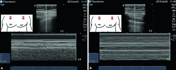
Abb 8 8 A B M Mode Am Rechten Hemithorax Normalbefund Weisse

Pneumothorax m motion mode.
Pneumothorax ultraschall m mode. Die sonographie eignet sich offenbar gut zur raschen pneumothorax diagnostik haben jenaer pneumologen festgestellt. Mit ultraschall erkennt man pneumothorax und infiltrate gut jena ner. Slide 3 of 3. Bei normalen verhältnissen entsteht unterhalb der pleuralinie ein granuliertes muster während im bereich der cutis subcutis und interkostalmuskulatur ein lineares muster entsteht seashore sign.
Ultrasound for detection of pneumothorax written by angela cirilli rebel em medical category. One way that we could represent the motion from lung sliding in a still image is to use m mode or motion mode and this is where you take a one dimensional line put it across the pleural line and look at it over time. Bei normalen verhältnissen entsteht unterhalb der pleuralinie ein granuliertes muster während im bereich der cutis subcutis und interkostalmuskulatur ein lineares muster entsteht seashore sign. Slide 1 of 3.
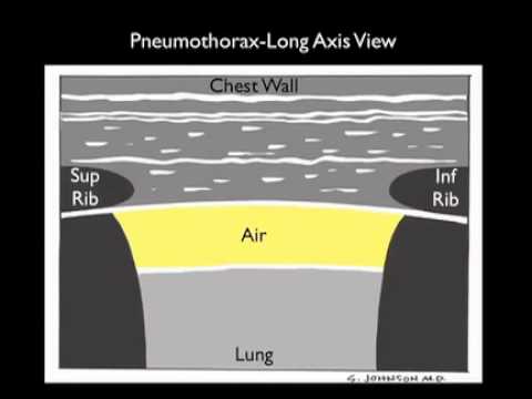
Thoracic and respiratory 12 comments typically the initial evaluation of blunt trauma patients involves a supine anteroposterior ap chest x ray cxr which has a poor sensitivity for the detection of pneumothorax ptx and has been reported as low as 20 48. Ein strahl wird durch das subkutane gewebe die interkostalmuskulatur pleura und lunge gelegt. Drücken der m taste zum starten des m modus. Betätigen des trackballs zum verschieben der achse.
Schmaler pleuraerguss pneumothorax. Slide 2 of 3. Im m mode wird ein strahl durch das subkutane gewebe die interkostalmuskulatur pleura und lunge gelegt. Bei vorliegen eines pneumothorax ist aufgrund der fehlenden bewegung im bereich der.
Beim m mode wird eine achse in das b bild gelegt und das signal das von den reflektierten ultraschallwellen dieser achse empfangen wird über die zeit aufgetragen. Eine achse wird im sono bild definiert und das signal. Bei einem pneumothorax lösen sich die beiden pleurablätter voneinander ab und das. Ultraschall gesteuerte punktion indiziert lungenarterienembolie.
Keilförmige herde zum hilus hin oft abgerundet meist dorsobasal. Normal motion seen below pleural line seashore sign continue narration. Dieses video enthält eine anleitung zur verwendung patientennaher ultraschalldarstellung und hochfreque. Die diagnose durch ultraschall ist eine neuere methode schnell zuverlässig und für einen kleinen pneumothorax mantelpneumothorax sensibler als die röntgenaufnahme.
Beim pneumothorax zeigt der m mode das barcode zeichen wohingegen beim normalbefund das seashore zeichen zu sehen ist.
Https Www Thieme Connect De Products Ebooks Pdf 10 1055 B 0034 15109 Pdf

Color And Power Doppler Sonography For Pneumothorax Detection
Http Www Degum De Fileadmin Dokumente Sektionen Chirurgie Vortr C3 A4ge Jahrestagung 2015 09 Breitkreutz Thorax Trier 150606 Pdf
Https Www Thieme Connect De Products Ebooks Pdf 10 1055 B 0034 15140 Pdf
Https Www Thieme Connect De Products Ebooks Pdf 10 1055 B 0039 168236 Pdf
Pdf Praklinische Notfallsonographie
How To Ultrasound For Pneumothorax Case Study Youtube

