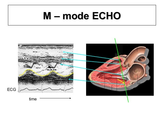
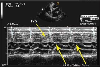
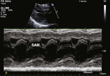
Sam M Mode Echo

M Mode Echo Principles And Classic Findings Wikidoc

Intracardiac M Mode In A Patient With Hypertrophic Obstructive

Systolic Anterior Motion Sam Sonopath

Theheart De Hocm Diagnose

Systolic Anterior Motion Sam Of The Mitral Valve Visualized By M
Dr Purvi Parwani On Twitter Going Back To The Basics Echofirst
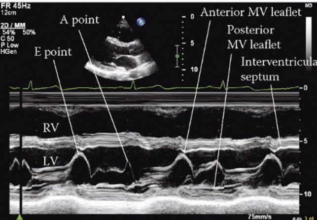
Here we will discuss how and where one should use the m mode.
Sam m mode echo. Es zeigt mit dem sam phänomen die einengung des ausflusstraktes durch di mitralklappe. Department of medicine division of cardiovascular disease clinical cardiology royal postgraduate medical school hammersmith hospital london w12 england. Vena contracta 6 mm 14. Im m mode von der glei chen anlotung aus.
Get free echo lectures 2 4 m mode. Sonografie oder sonographie auch echografie und ultraschalluntersuchung oder umgangssprachlich ultraschall genannt ist ein bildgebendes verfahren mit anwendung von ultraschall zur untersuchung von organischem gewebe in der medizin und veterinärmedizin sowie von technischen strukturen. Supported by the deutsche forschungsgemeinschaft during part of this study. Insuffizienzjet 50 des lvot diameters bzw.
Die obstruktion in ruhe ist nur leicht mit werten um 30 mmhg. Although the interrogating ultrasound can be tilted to visu alize different structures and their anatomic relations it may miss other. Das ultraschallbild wird auch sonogramm genannt. Hochfrequentes diastolisches flattern des vorderen mitralklappensegels 12.
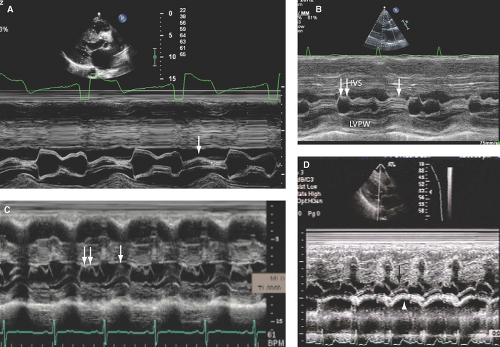
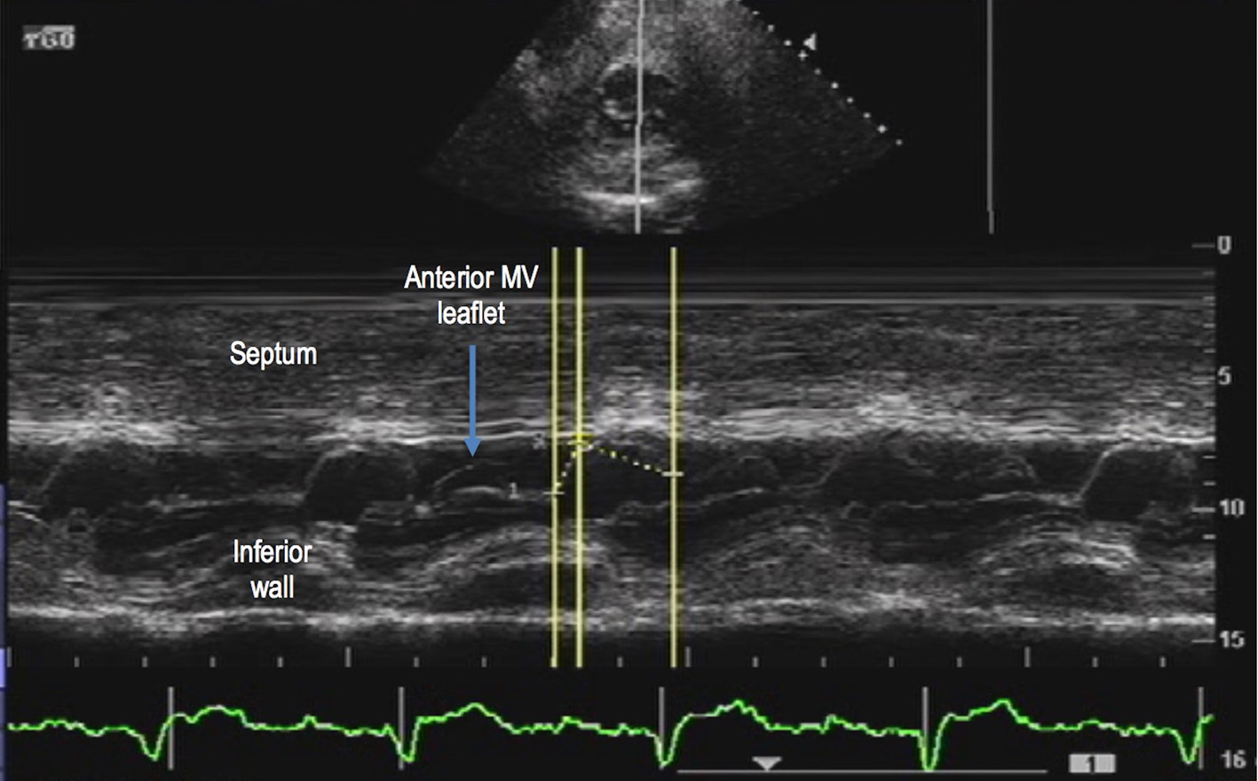
Echocardiographic predictors of left ventricular outflow tract obstruction and systolic anterior motion of the mitral valve after mitral valve reconstruction for myxomatous valve disease. The 2d reference image will help you identify the individual structures you see on the m mode. Die sam systolic anterior movement der mitralklappe kann deutlich erkannt werden. In chapter 1 we described the basic principles of m mode imaging.
Mittels cw doppler erfasste geschwindigkeiten im lvot. In addition you will learn about the structures that can be visualized and how the measurements should be performed. Journal of the american college of cardiology 34 7 2096 lp 2104. Darstellung der mitraklappe mit dem sam phänomen während der systole bei typischer hocm mit hilfe der belastungsechokardiografie echo untersuchung unter belastung auf einem fahrradergometer in schräglage wird die auswirkung der erkrankung unter belastung zuverlässig.
Der lvot oberfläche kurze achse 15. Hochfrequentes diastolisches flattern des interventrikulären septums farb doppler 13.
Systolic Anterior Motion Of Mitral Valve Sam
Hypertrophic Cardiomyopathy Thoracic Key
Echocardiography Clinical Gate
Hypertrophic Cardiomyopathy Anesthesia Key

Acep American College Of Emergency Physicians

Physicians Academy Jan 2018 Volume 12 Number 1 Article 1
Systolic Anterior Motion Feline Echocardiography Vetgirl Vet Ce Blog
Systolic Anterior Motion Of Mitral Valve Subchordal Apparatus A

